About this image
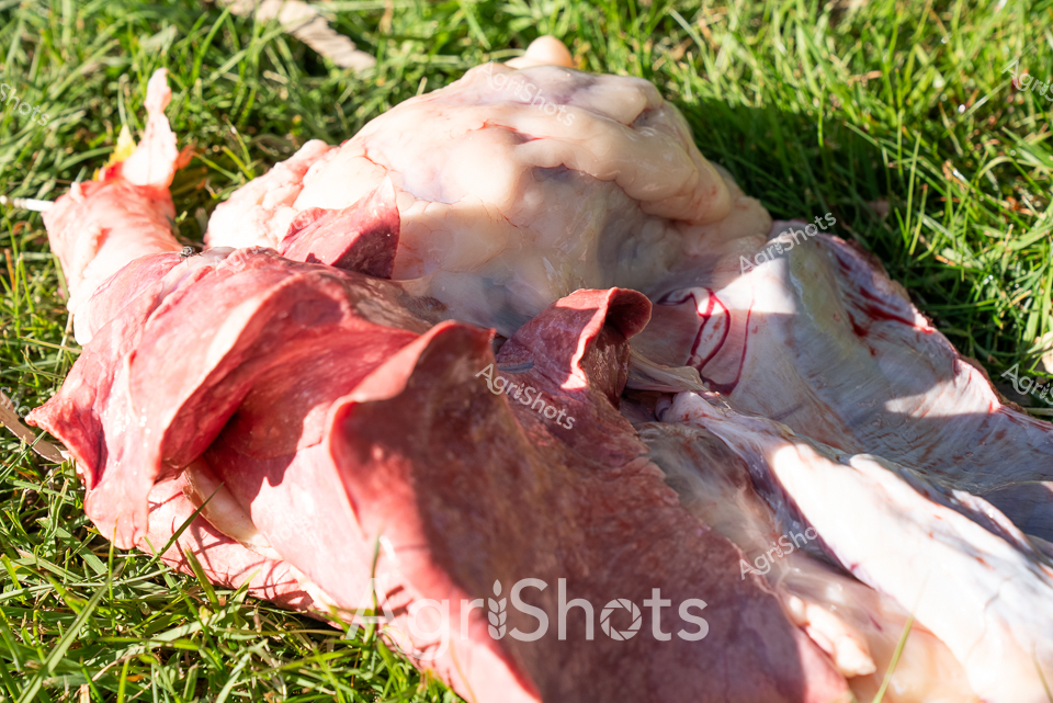
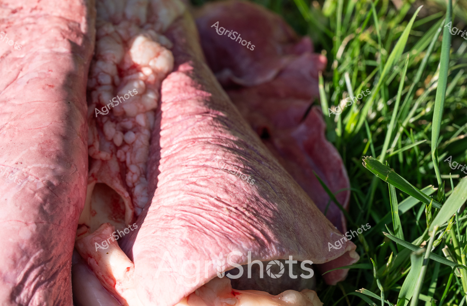
Fresh meat cuts reveal deep red and pale pink tissues, highlighting the transition from livestock to food production. Anatomical segments demonstrate the professional meat processing techniques used in Australian agricultural supply chains. Sunlight illuminates the textural details of the butchered meat, casting sharp shadows across the grass and revealing the precise cutting and preparation methods. The graphic close-up provides a clinical view of livestock processing, representing an essential stage in meat production and agricultural food systems.
Similar media
 Image
Image
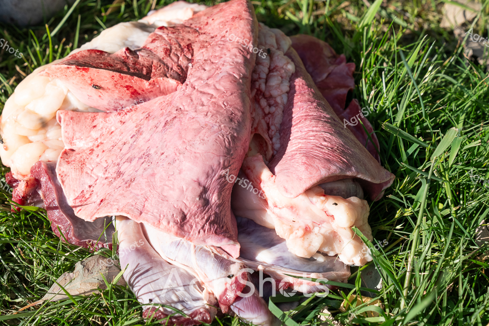
Raw Ovine Anatomical Study: Thoracic Dissection Pale pink and deep crimson lung tissue sprawls across verdant grass, presenting an intimate anatomical landscape of ovine respiratory anatomy. The delicate, textured organs display intricate structural details - lobes, vessels, and membrane variations revealing the complex internal architecture of a sheep's pulmonary system. Soft tissue transitions from pale blush to deeper carmine hues, highlighting the organic contours and subtle variations in tissue density. Sharp blades of grass provide a stark, living green counterpoint to the exposed biological specimen, creating a visceral contrast between vibrant vegetation and the vulnerable internal anatomy. This forensic-like examination offers a clinical yet poetic glimpse into the biological mechanics of livestock, capturing the raw intersection of scientific observation and agricultural practice. The precise arrangement suggests methodical veterinary or agricultural research, documenting the physiological details critical to understanding animal health and livestock management.
 Image
Image
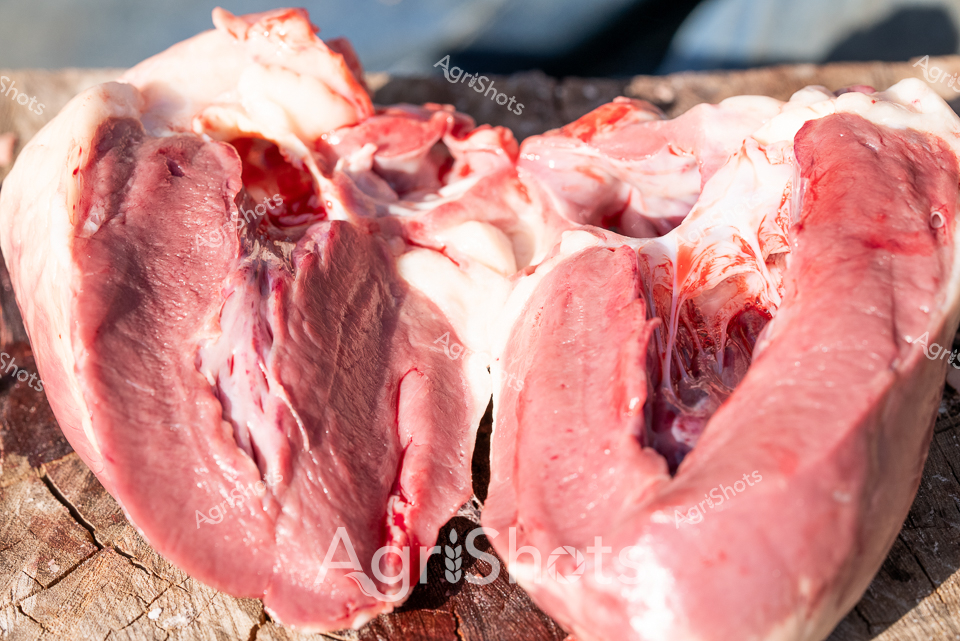
Raw Ovine Cardiac Anatomy: Veterinary Pathology Documentation Freshly sectioned sheep heart rests on weathered timber, revealing intricate internal anatomical structures in vivid crimson and pale pink hues. Precise surgical dissection splits the muscular organ longitudinally, exposing complex chambers, vessels, and tissue layers with forensic clarity. Deep red muscle tissue contrasts against lighter membrane surfaces, capturing the extraordinary biological engineering of ovine cardiovascular systems. Forensic veterinary pathologists and agricultural researchers rely on such detailed post-mortem examinations to understand livestock health, genetic conditions, and potential disease transmission. The raw, unfiltered presentation speaks to scientific documentation's critical role in maintaining agricultural biosecurity and animal welfare standards. Captured with clinical precision, this image represents the intersection of veterinary science, agricultural research, and systematic anatomical investigation.
 Image
Image
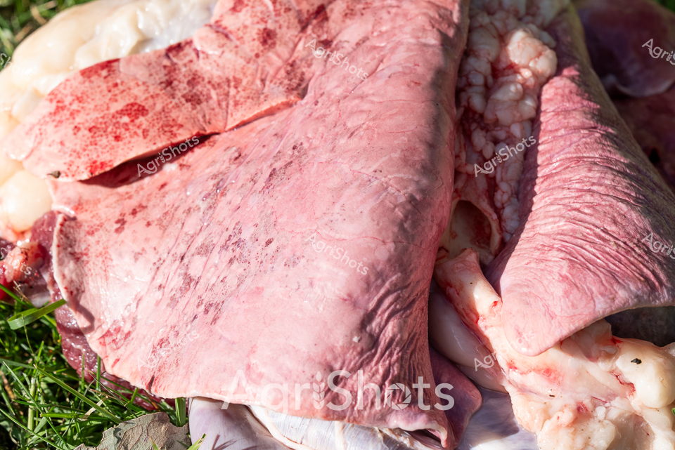
Raw, freshly harvested sheep lungs rest on a bed of green grass, revealing the intricate anatomical details of this vital organ. The pale pink tissue displays a delicate, textured surface marked with subtle variations in color and translucent membrane layers. Deep crimson spots and residual blood highlight the recent harvest, capturing the clinical precision of veterinary or agricultural inspection processes. Soft, folded surfaces and varying tones of pink and white demonstrate the complex internal structure, with subtle veining and tissue connections visible. The surrounding grass provides a stark, natural contrast to the clinical, raw biological specimen, suggesting a farm setting where animal health and livestock management are carefully monitored. This close-up reveals the meticulous attention to detail in agricultural practices, showcasing the unfiltered reality of livestock processing and veterinary examination.
 Image
Image
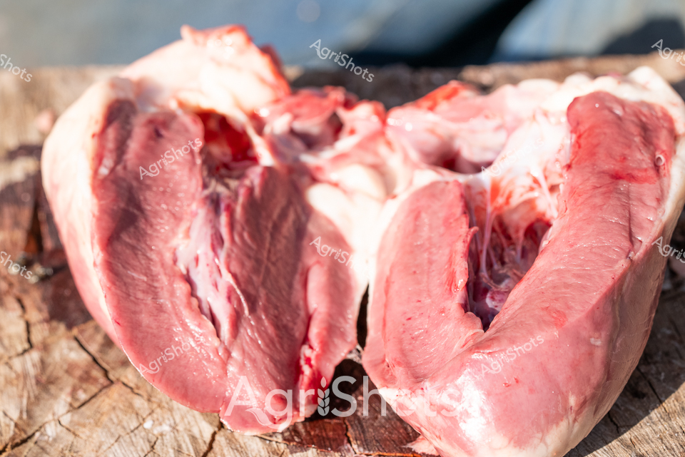
Raw Anatomical Precision: Sheep Visceral Examination A freshly sectioned ovine heart lies split open on a weathered wooden surface, revealing intricate internal anatomical structures with clinical clarity. Deep crimson muscle tissue displays rich, dense textures highlighting the complex cardiovascular system, with chambers and arterial pathways exposed in precise detail. The vivid pink and dark red tones contrast sharply against the rough-hewn timber backdrop, suggesting a professional veterinary or agricultural research environment. Subtle moisture glistens across the tissue surface, indicating a recent dissection under controlled, scientific conditions. This forensic-grade documentation captures the intimate biological architecture of livestock cardiovascular anatomy, serving critical purposes in animal health research, veterinary training, and agricultural science. The meticulous cross-section provides researchers and agricultural professionals an unobstructed view of physiological structures essential to understanding sheep health and biological systems.
 Image
Image
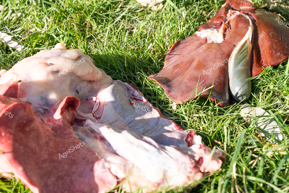
Raw pastoral pathology unfolds across a verdant grass field - a post-mortem examination of sheep viscera illuminates agricultural veterinary science. Pale pink and deep crimson organ tissues sprawl across bright green grass, revealing intricate anatomical details of lungs, liver, and connective membranes. Sunlight casts sharp shadows, highlighting the delicate textures and organic contours of the dissected biological specimens. This forensic agricultural scene captures the critical diagnostic processes farmers and veterinarians employ to understand livestock health, disease transmission, and biological conditions affecting sheep populations. The clinical yet naturalistic arrangement speaks to the intersection of scientific methodology and rural animal husbandry practices. Each exposed tissue fragment represents a potential window into understanding complex physiological systems that underpin livestock management and agricultural sustainability.
 Image
Image
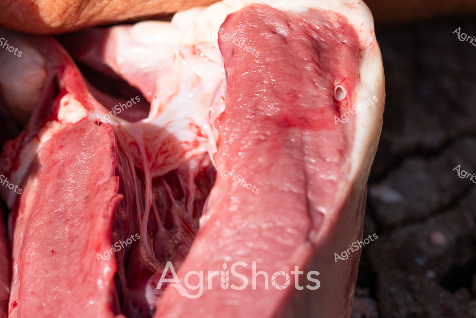
Dissection reveals the intricate internal anatomy of a fresh sheep carcass, with raw muscular tissue displaying rich crimson and pale pink hues. Dense muscle fibers and connective tissue intersect in complex layers, demonstrating the biological structure beneath the animal's exterior. The precise cross-section exposes the dense musculature and structural composition, highlighting the professional veterinary or agricultural research context of this detailed anatomical examination. Textural variations range from smooth, glistening membrane surfaces to the deep, striated patterns of muscle tissue, capturing the biological complexity of ovine physiology. This forensic-level documentation provides critical insights into livestock health, meat quality assessment, and veterinary diagnostic procedures. The high-resolution image underscores the scientific precision required in agricultural research and animal health monitoring.
 Image
Image
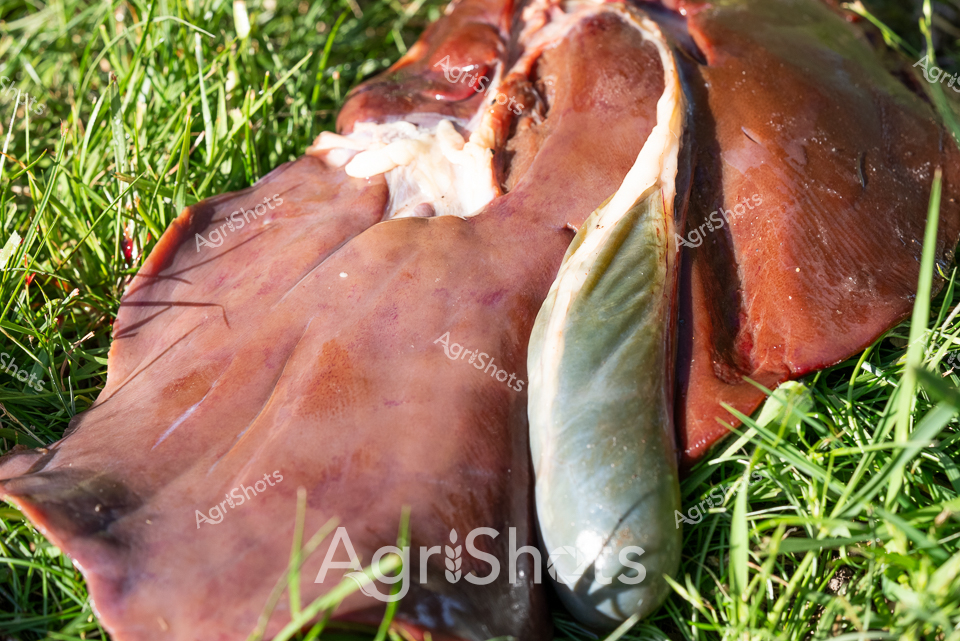
Fresh Ovine Dissection: Anatomical Research Reddish-pink sheep viscera sprawl across vibrant green grass, revealing intricate internal anatomy during a precise post-mortem examination. The raw biological tissue displays rich crimson and pale cream tones, with delicate organic structures exposed against the verdant pasture backdrop. This forensic agricultural investigation captures the methodical process of livestock health monitoring and veterinary research, highlighting the scientific approach to animal husbandry. Each carefully separated organ demonstrates the meticulous nature of agricultural science, where understanding animal physiology is crucial to maintaining flock health and productivity. The high-resolution close-up provides an unflinching glimpse into the complex biological systems that underpin successful sheep farming, presenting a stark yet scientifically fascinating view of livestock management.
 Image
Image
Raw, visceral anatomical study of sheep respiratory system, displaying pale pink lung tissue against verdant grass. Detailed cross-section reveals intricate internal structures, with delicate alveolar clusters and textured pulmonary membranes. The pale organic matter contrasts sharply with bright green grass blades, creating a clinical yet naturalistic agricultural pathology scene. Precise dissection exposes the complex biological architecture, suggesting veterinary research or livestock health monitoring processes. The graphic anatomical detail speaks to the meticulous scientific approach fundamental to modern agricultural practice.